Seeing DiMEs With a Magnetic SNOM
A new tool combines a cryogenic nanoscope with a strong magnetic field to visualize quantum particles at sub-nanometer scales.

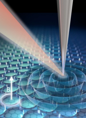
Scientists at Stony Brook University, Columbia University, and the University of California San Diego have developed a new tool to image quantum particles called Dirac magnetoexcitons (DiMEs) at infrared frequencies for the first time. The tool, described in a paper published recently in Nature Nanotechnology, combines a cryogenic scattering-type scanning near-field optical microscopy (SNOM) with high magnetic fields.
SNOM is a widely used technique to study quantum particles that are smaller than the diffraction limit of light. To fully probe the properties of these particles, however, researchers need the microscopes to work at cryogenic temperatures and in the presence of strong magnetic fields—technical challenges that the team overcame by using a special type of atomic force microscope tip called an Akiyama probe along with careful, compact positioning inside of a vacuum chamber.
With their cryogenic magneto-SNOM, the researchers were able to directly visualize and map the behavior of DiMEs in graphene. DiMEs are collective excitations of electrons that occur when a magnetic field is applied. “Imaging the DiMEs directly at their native length scale is an exciting discovery because they had previously only been observed using far-field optics, which cannot measure the nanoscale properties that would make DiMEs useful in different applications,” explained co-author Mengkun Liu, a physicist at Stony Brook University and Brookhaven National Laboratory who collaborates with Columbia as part of the Department of Energy-funded EFRC on Programmable Quantum Materials. Those applications include high-speed optical communication and sensing, and as a platform for quantum computing.
The results were a surprise to the team. “For the first non-test experiment done with the magneto-SNOM, we chose to look at the simplest and most widely studied two-dimensional material: graphene. We did the first measurement with no expectations of how the magnetic field would change the measurements,” said first author Michael Dapolito, who was completing his master’s degree at Stony Brook while the tool was developed and is now a Physics PhD student at Columbia.
It was late, around 11:00 PM, Dapolito recalled, when they got their first results. At zero magnetic field, the graphene was completely transparent. Then Liu decided to go straight to the max and applied a 7 Tesla magnetic field. The graphene sheet lit up, with clear fringes along the edges. “This was massively different from the zero-field case, completely unexpected, and quite exciting to see—especially at our nanoscale spatial resolution,” he said. This nanoscale imaging revealed how the DiMEs form standing waves in optical signals and drive unique photocurrents along the perimeter of the sample.
“Electrons in a magnetic field do strange things that we haven’t seen before, because the resolution of existing tools that are compatible with magnetic fields hasn’t been good enough,” said co-author and Columbia physicist Dmitri Basov. “These results should be the first of many with this monumental new tool.”
The researchers plan to continue improving the cryogenic magneto-SNOM and use it to study different quantum materials and phenomena, including exotic quasiparticles, superconductors, and ultrafast electron dynamics–providing new insights into quantum physics at sub-nanometer scales.
Read More: Michael Dapolito et al. Infrared nano-imaging of Dirac magnetoexcitons in graphene. Nature Nanotechnology (2023). DOI: 10.1038/s41565-023-01488-y
Competing interests: Four of the authors have a patent pending related to the design of the magneto-SNOM. The other authors declare no competing interests.
